
By Sil, founder of Spores Worldwide · prepared in our Ottawa lab · Updated October 2026 · For microscopy and taxonomy research only.
Mushroom spores are the microscopic reproductive cells that mushrooms release, much as plants release seeds. Each spore is a single cell. Mushrooms make them on their gills and drop them into the air, and when enough land in one place they form a visible spore print. Under a microscope, spores have a size, shape and colour that are characteristic of their species, which is why mycologists study them.
This guide explains what spores are, how a mushroom makes and releases them, and what they look like on a slide. Spores Worldwide supplies Psilocybe cubensis spores for microscopy and taxonomy research only.
Spores are single cells, not seeds
Spores and seeds do a similar job: they spread an organism to new places. But they’re built very differently. A seed contains a developing embryo, while a spore is unicellular: one cell (Wikipedia, Spore).
Mushrooms belong to the Basidiomycota, and their spores are called basidiospores. Each one typically carries a single haploid nucleus, the product of meiosis, so it holds one set of the parent’s genetic material rather than two (Wikipedia, Basidiospore). That’s also why spores from the same mushroom aren’t identical copies of it.
Where spores are made
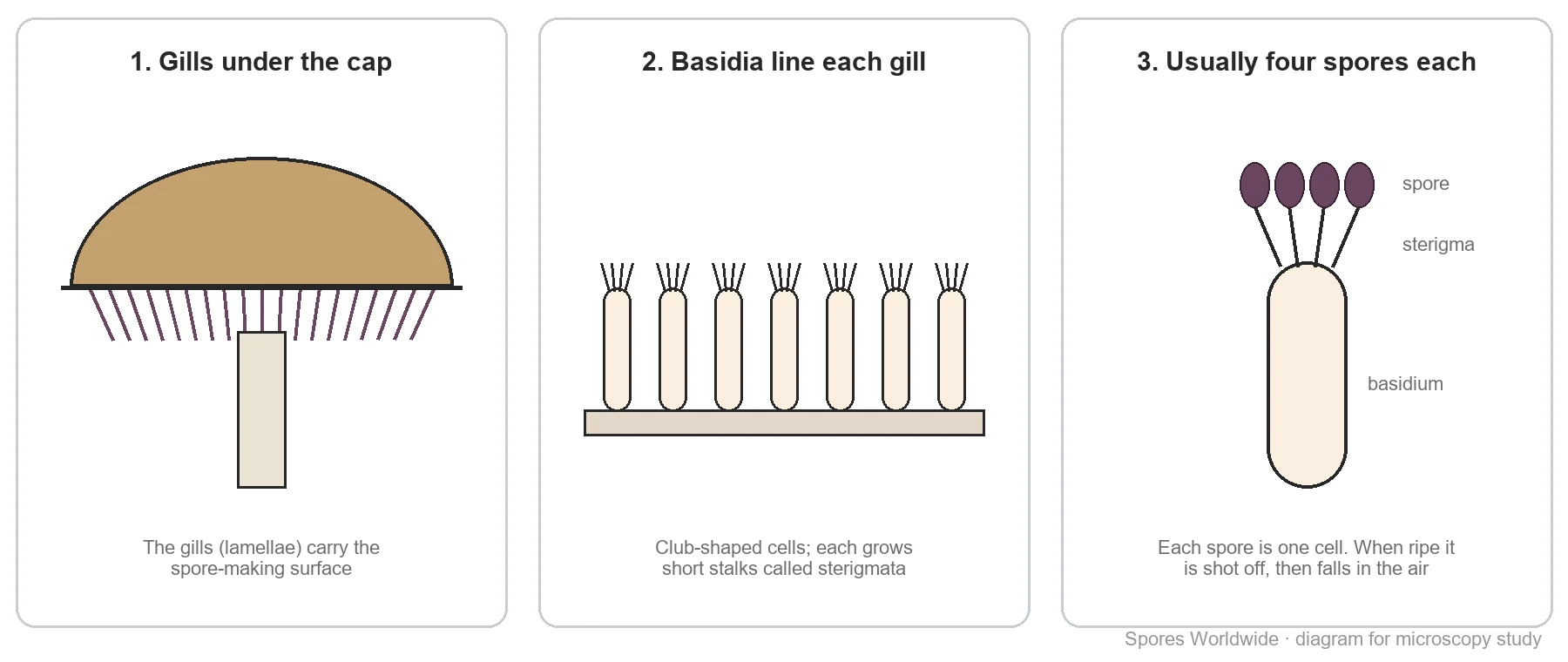
On a gilled mushroom like Psilocybe cubensis, spores are made on the gills (lamellae) under the cap. Other mushrooms use pores or teeth instead, but the principle is the same.
The surface of each gill is lined with club-shaped cells called basidia. Each basidium grows small stalks called sterigmata, and a spore develops at the tip of each one. Typically there are four spores per basidium, although the number can range from one to eight. P. cubensis basidia are usually 4-spored, sometimes 2- or 3-spored (Wikipedia, Psilocybe cubensis).
How spores are released
Mushroom spores aren’t simply shaken loose. When a spore is ripe, a tiny droplet of water called Buller’s drop forms near its base. In a fraction of a second the droplet merges with a film of water on the spore, and the sudden shift launches the spore a short distance off the gill (Wikipedia, Ballistospore). It then falls out from between the gills and is carried away by air currents.
If the cap sits still over a surface, the falling spores pile up directly beneath the gills. That’s how a spore print forms.
Spore prints: spores you can see
A spore print is the powdery deposit left when spores fall from a cap onto a surface underneath. Its colour is an important identification feature: prints are usually white to cream, black, or shades of red, purple or brown. But no mushroom can be identified from its print alone; it’s one feature among several (Wikipedia, Spore print). The colour comes from the spore wall, so the print and the individual spores tell the same story.
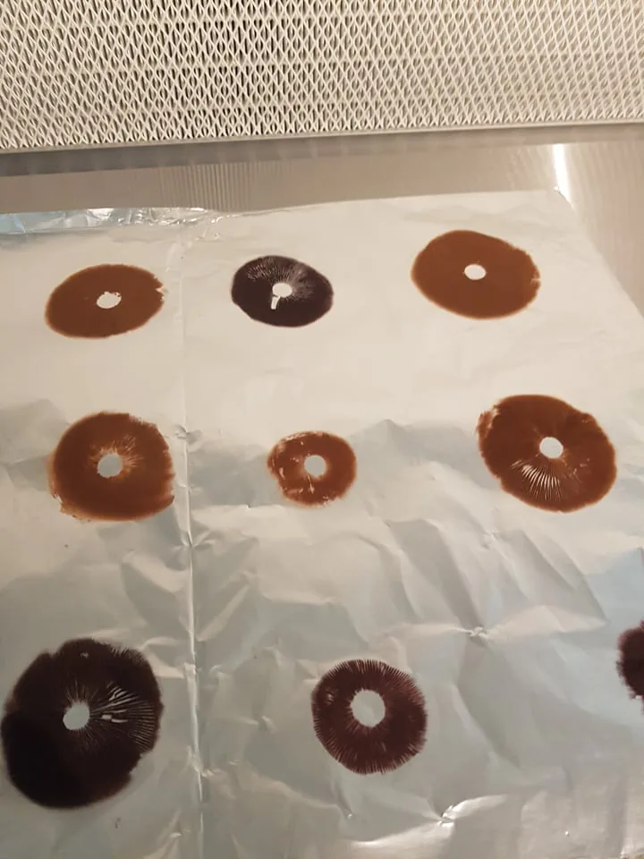
Psilocybe cubensis prints are purple to dark purple-brown. For how prints are made and kept, see How to make a spore print.
What spores look like under a microscope
At 100× spores are fine specks; at 400× you can see their outline and colour; at 1000× with oil immersion you can measure them. Mycologists record four things:
- Size, in micrometres (µm; 1 µm is a thousandth of a millimetre), measured with a calibrated eyepiece reticle.
- Shape: round, elliptical, almond-shaped, angular and so on.
- Colour, which matches the spore print.
- Surface and wall: smooth or ornamented, thin or thick.
Two mushrooms that share the nickname “Blue Meanie” show how useful this is:
| Psilocybe cubensis | Panaeolus cyanescens | |
|---|---|---|
| Spore print | Purple | Black |
| Spore size | 11.5–17.3 × 8–11.5 µm | 12–15 × 7–11 µm |
| Spore shape | Sub-ellipsoid | Elliptical, smooth, opaque |
Sources: Wikipedia, Psilocybe cubensis and Panaeolus cyanescens.
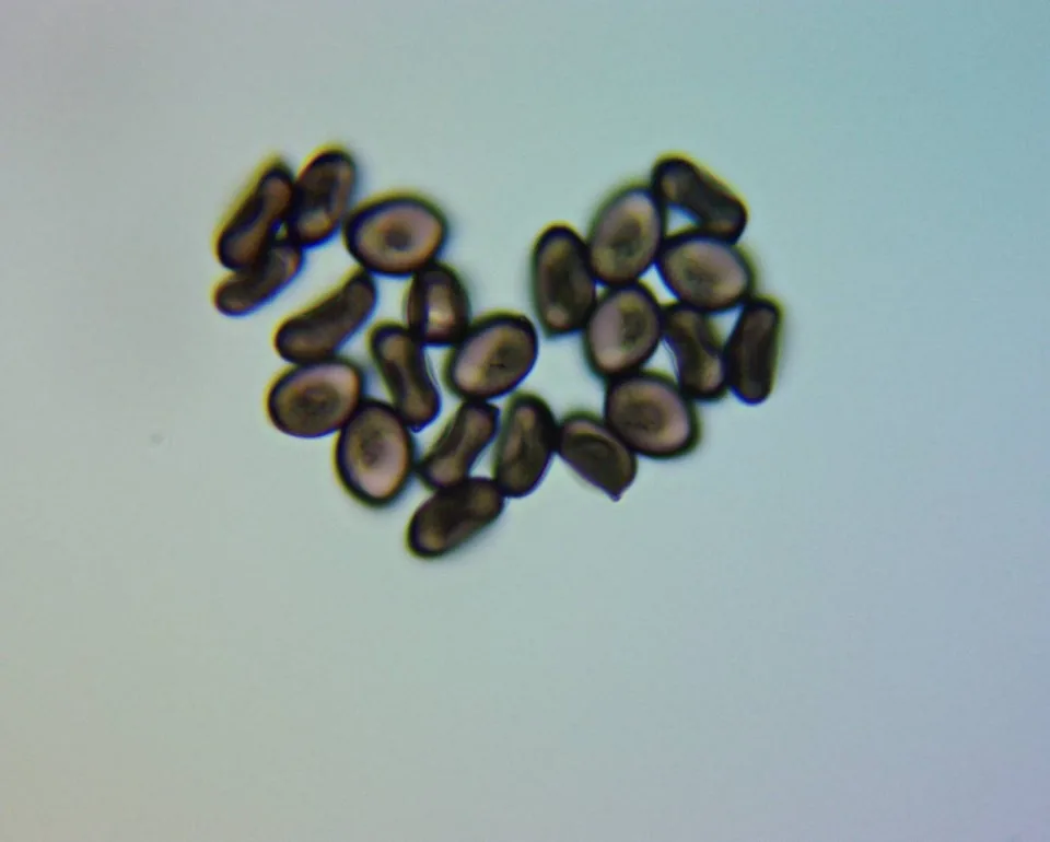
To prepare your own slide, follow How to use a spore syringe for microscopy.
Spore syringes, prints and swabs
Spores reach a microscopist in one of three formats:
- Spore syringes: spores suspended in sterile water. The easiest way to put a drop on a slide.
- Spore prints: the dry deposit on foil. Best for print-colour study and long-term storage.
- Spore swabs: spores collected on a sterile swab, a compact alternative to a print.
All three hold the same kind of single-celled spores described above. To keep them in good condition, see How to store spore syringes, prints and swabs. Whether you can own spores depends on where you live; see Are mushroom spores legal?.
Related reading
- How to use a spore syringe for microscopy
- How to make a spore print
- Golden Teacher spores: strain profile
- Penis Envy spores: the PE family
Frequently asked questions
What are mushroom spores?
Microscopic, single-celled reproductive units that mushrooms release to spread. Gilled mushrooms make them on the gills, usually four on each basidium.
Are mushroom spores the same as seeds?
No. They do a similar job, but a seed contains a developing embryo, while a spore is a single cell.
How big are mushroom spores?
It varies by species. Psilocybe cubensis spores are 11.5–17.3 × 8–11.5 µm (a micrometre is a thousandth of a millimetre), so you need a microscope at 400x or more to see their shape.
What colour are mushroom spores?
It depends on the species: spore prints are usually white to cream, black, or shades of red, purple or brown. Psilocybe cubensis prints are purple to dark purple-brown.
How do you look at mushroom spores?
Put a small drop from a spore syringe (or a little print scraped into sterile water) on a clean slide, add a cover slip, find the spores at 100x, and study them at 400x to 1000x.